
Treating Infections, Injuries and Disease in a Horse's Eye
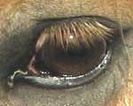 The globe of the horse's eye, the eyeball, is an extremely
specialised structure, closely coordinated with the central nervous system
The globe of the horse's eye, the eyeball, is an extremely
specialised structure, closely coordinated with the central nervous system
The tissues surrounding a horse or pony's eye, the eyelids and membranes, are closely associated with the skin of the head.
Horses have a very wide angle of vision; the position of the eyes high on the sides of the head allows a horse to see what is going on at its shoulder and down its flanks on both sides, without turning its head from side to side.
Good eyesight is essential for the accurate evaluation of jumps and obstacles: a partially sighted heavy horse may work well without significant disability, but an event horse or show jumper with only limited vision is dangerous.
TREATING TRAUMATIC INJURY AND INFECTIONS OF A HORSE'S EYE
Horse's eyelids are frequently torn by wire, ripped by bites from other horses or shredded by a kick to the eye.
Because of the great importance of conserving the conformation of the eyelids, so that the drainage of the tears is preserved, all eyelid injuries should be taken seriously.
A small gash on the side of the face away from the eye may be left to heal without suturing, but such a gash in an eyelid may need careful surgical repair.
In this case it is best to call your vet who will check that both the globe of the eye and the underlying bone of the orbit have not been damaged.
These may require attention before the eyelid wound is tackled.
For the repair of eyelids small, non irritating sutures are used. The vet will attempt to reconstruct the margin of the lid; skin can be pulled from around the horse or pony's eye to help make up deficits using a 'sliding graft' technique.
Medication of a damaged eye or eyelid can be difficult. The vet will sometimes sew into the eyelid a very fine irrigating cannula which is placed through the upper lid and attached across the top of the head and down the neck.
Using this system, antibiotics and other medications can be flushed into the eye by pumping them from a syringe at the horse's withers, avoiding all the restraint and wrestling necessary to insert cream or ointment into a painful eye in an adult horse.
EQUINE CONJUNCTIVITIS
Conjunctivitis is common in horses.
The condition shows as a discharging eye, sometimes with a swollen eyelid; the horse will keep the affected eye closed, especially in bright light.
Some cases begin with an allergy - certain horses are affected seasonally by certain pollens or dusts, but most cases have an element of infection involved which turns the clear fluids of the normal eye into thicker, more mucoid discharges or even into pus.
Two further factors complicate the condition; first, any discharge from the eye or overspill of tears will attract flies which cause more irritation and introduce more contamination, whilst secondly, the thicker fluid in the eye may block the drainage system of the horse's tear ducts, resulting in total overspill down the side of the face.
Use clean water only to wipe away the muck from the side of the face and avoid cotton wool or soft tissues, which may shed fibres and exacerbate the irritation.
Any discharging or swollen eye should be checked by an equine veterinary surgeon.
PARALYSIS OF A HORSE'S EYELID
Swelling and closure of the eyelids because of infection, should not be confused with drooping of the eyelid due to paralysis.
The lids may be paralysed, usually on one side because of damage to a nerve elsewhere; the paralysis may be accompanied by retraction of the horse's eye, sweating on one side of the face or other signs depending on the exact location of the insult to the nerve.
BLOCKED TEAR DUCTS
This may occur either because of a primary problem within the drainage channel, which runs from the inside margin of the eye, through the sinuses and drains into the nose, or because of conjunctivitis which has thickened the fluid in the eye and clogged the system.
The tear ducts can be flushed by the vet quite easily, as the exit orifice is accessible within the nostril.
TUMOURS
The horse's eyelids are a prime site for the development of cancerous growths, either benign or malignant.
These may initially appear as little bumps below the skin of the eyelid or as small warts' on the margins of the lid.
They should never be ignored.
Only one type, the juvenile virus papilloma can safely be left untreated; these occur in young horses and appear as a large crop or outbreak of warts on the face, especially the muzzle and around the eyes.
They will disappear within a few weeks.
All other eyelid tumours are potentially serious and should be examined by a veterinary surgeon.
THE THIRD EYELID
Across the inside angle of the eye is a shutter of moist tissue, the nictitating membrane, or third eyelid. This structure is usually retracted and barely visible, but in one or two conditions becomes more noticeable.
In cases of infection or allergy, the lymphoid tissue on the surface of the membrane may become enlarged and inflammed. This results in the appearance of a bubbly pink blob in the corner of the eye. This should be checked - infection will not be serious, but occasionally this is the first sign of a cancerous growth.
The obvious protrusion of both nictitating membranes occurs early in the course of Tetanus, another very serious condition.
ENTROPION IN FOALS - INWARD ROLLING EYELID
Foals are occasionally born with an eyelid which rolls inwards, so that the front of the eye is in contact with the eyelashes and hair, rather than with the moist membrane of the conjunctiva.
This soon causes inflammation, making the eye look weepy and probably partly closed.
Mild cases of entropion will unroll in the first few weeks of life as the foal grows and the eyelids turn up, so that the conformation of the eyelids ends up perfectly normal.
During this period the eye can be protected with regular application of ointment.
In some cases, however, the deformity is severe enough to cause an ulceration of the front of the eye.
If this occurs, or if the vet fears that there is a risk of this occurring, surgical correction of the blemish can be carried out.
Surgery is simple, but needs to be precise. Under general anaesthetic, a crescent shaped portion of skin is removed from underneath the margin of the eyelid (lower lids are more frequently affected).
When the edges of this cosmetic wound are brought together, the tension rolls the eyelid outwards and corrects the entropion.
Removing too much tissue creates an eyelid which folds outwards leading to other problems; removing too little does not correct the entropion in the foal's eye.
HORSE CARE ADVICE AND EQUESTRIAN ARTICLES
| Feeding Linseed to Horses | Causes of horse nosebleeds | Redworm | Veteran Horse Care | Devils Claw | Equine kissing spines | Horse Colic | Treating Cracked Heels | Removing Bot Fly Eggs | Feeding Haylage | Soaking Hay | Arnica for bruised soles | Rain Scald | Horse livery Wiltshire | Sweet Itch | Insurance | Sidebone | Eating Mud | Feeding Carrots and Apples | Hoof Care | Ragwort | Aubiose |